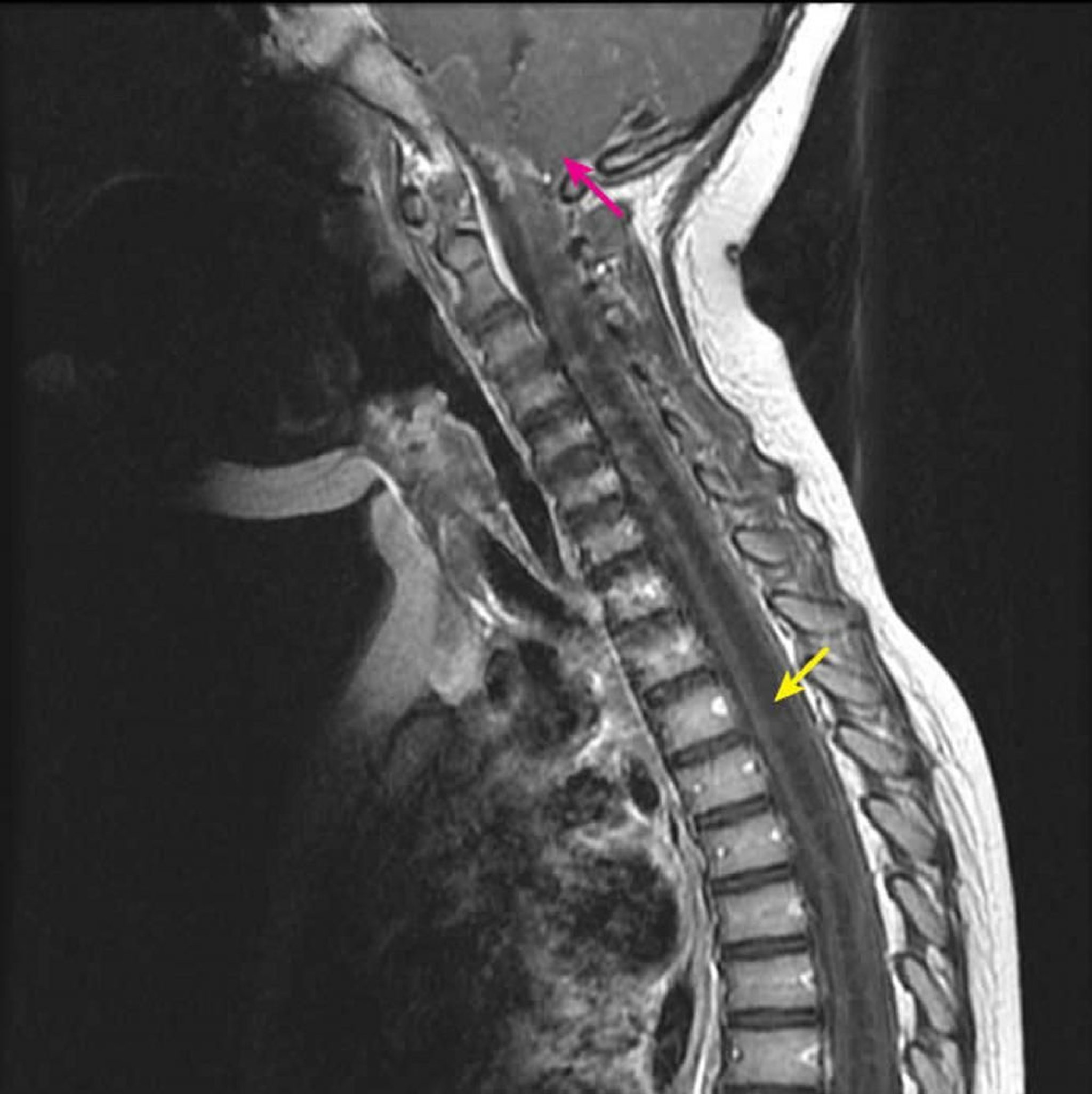
颈胸段脊髓空洞积水症
矢状位增强后 T1 加权 MRI 显示一个大的 T1 低信号髓内脊髓腔,符合脊髓空洞特征(黄色箭头)。在已知 Chiari I 畸形(粉红色箭头)的儿童中,它从 C2-3 颈椎水平向下延伸。这一发现被定义为脊髓空洞积水症,因为脊髓空洞症(偏心脊髓腔)无法通过影像学与脊髓积水(中央脊髓腔)区分开来。
Courtesy of John Tsiouris, MD, Division of Neuroradiology, New York–Presbyterian Hospital/Weill Cornell Medical Center.
在这些主题中